Effect of linear index mismatch and near fields :
Nonlinear optical microscopes allow for label-free, molecule-specific, non-destructive imaging of biological processes in cells and tissues. Two such techniques are stimulated Raman scattering (SRS) microscopy and coherent anti-Stokes Raman scattering (CARS) microscopy, which allow for video-rate and hyperspectral imaging via molecular Raman resonances.
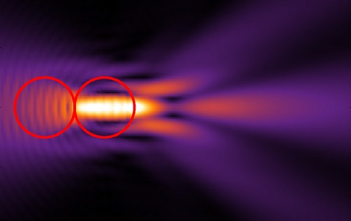
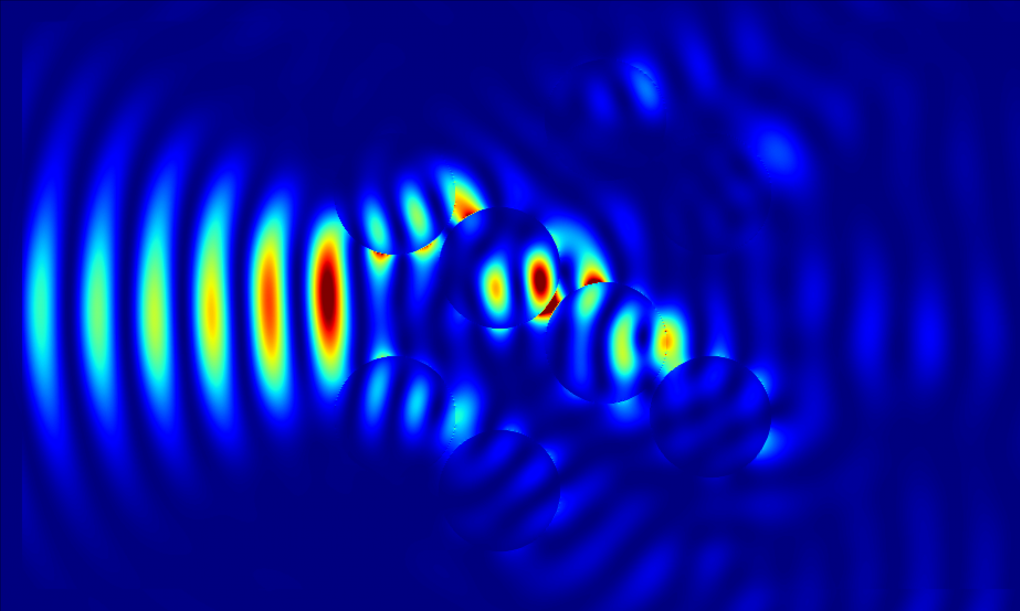
We show in Optics Express (2016) that an inhomogeneous linear refractive index profile, such as that occurring in biological tissues, is shown to significantly alter SRS and CARS images. In fact, near-field enhancement and microlensing can increase an object’s perceived molecular density by almost an order of magnitude and can change its perceived position by up to 1.0 μm. Furthermore false CARS and AM-SRS signals can be observed in the absence of Raman active media.
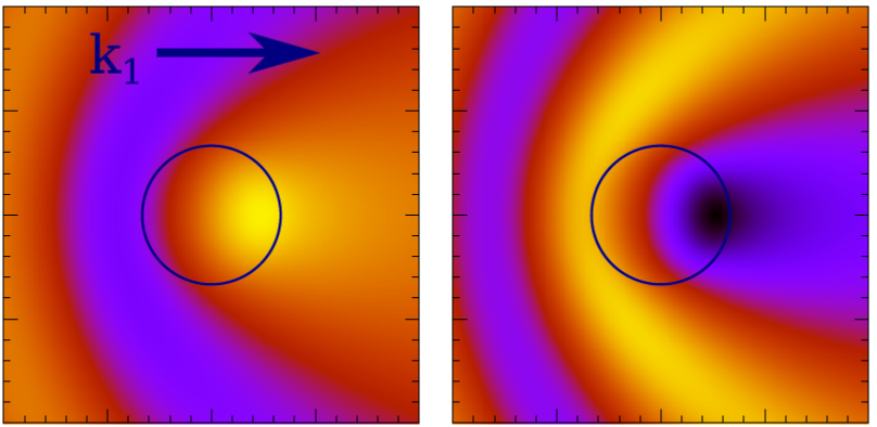
Nonlinear optical imaging using backward propagating signals is very sensitive to the location of objects along the laser axis, and thus are used to image very small features. In our Optics Letters (2018), we show both theoretically and experimentally that the inhomogeneous refractive index in tendon creates hot or cold spots in the constituent collagen fibrils, which can change the forward to backward signal ratio of the second harmonic generation (SHG) signal by up to 25%. This is caused by the extreme sensitivity of the backward signal on phase matching, which alters the destructive interference condition.