Velocity Map Imaging
Velocity map imaging (VMI) is a type of charged particle mass spectrometry. Time of flight spectrometers usually map ion mass/charge ratio to time. VMI spectrometers map ion velocity to position on a detector. This feature makes VMI a high-resolution ion imaging technique capable of determining highly differential information about the release direction and speed of ions, following a photophysical process.
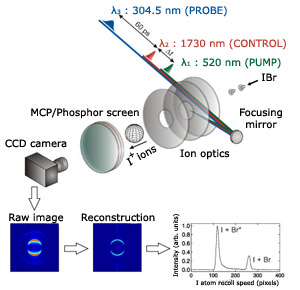
The approach is schematically illustrated in Figure 1. Neutral products of a photodissociation, reactive event, or the neutral electronically excited molecules are ionized by a probe laser. The created ions or photoelectrons are created in the spherical volume of the laser focus, and these spheres then expand according to the kinetic energy of the charged particles. After a certain time, fast charged particles (high kinetic energy) will be on the surface of a large sphere (fast expansion), and slow charged particles(low kinetic energy) on the surface of a much smaller sphere (slower expansion). By using an electrostatic lens, titled Ion optics in Figure 1, we project these nestled 3D-Spheres on a 2D-Detector – and by applying the appropriate voltages on the ion optics we can keep these nestled 3D-spheres in focus. The detector consists of 2 microchannel plates (signal amplification) and a phosphor screen, which will emit light when a charged particle hits the 2D-detector. A CCD camera captures the 2-D image which can then be used to reconstruct the 3-D velocity distribution. The distribution contains not only all the the kinetic energy of the charged particle, but also its angular distribution, which can provide a deeper insight in the observed dynamics. The use of ion optics eliminates the spatial blurring of the image due to finite size of molecular beam, allowing velocity resolution on the order of 1% or better. VMI allows detecting multiple events and the detection efficiency is extremely high. It is a powerful, versatile experimental technique. See Figure 3 for images of our VMI apparatus.
Laser systems
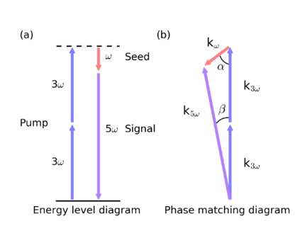
We use a 1KHz 35 fs Ti:Sa Laser system to generate probe and pump laser at different colors for our time-resolved experiments. Not only do we use the standard harmonics of 800 nm generated in BBO crystals as pump or probe (400, 266 and 200 nm), but we also recently implemented a scheme for the generation of VUV radiation, used either as a high-energetic pump or probe. VUV generation is based on the principle of non-collinear differenece frequency four-wave mixing (see Figure 2): 266 nm and 800 nm are focused under a small angle into a vacuum box filled with roughly 30 mtorr of Ar, and the created 160 nm is then seperated and focused into the machine using a set of dielectric mirrors. Using the output of an OPA as the seed, we recently implemented tunable VUV, which allows us an even broader range of possible experiments.
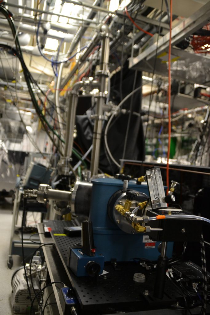
VMI-apparatus 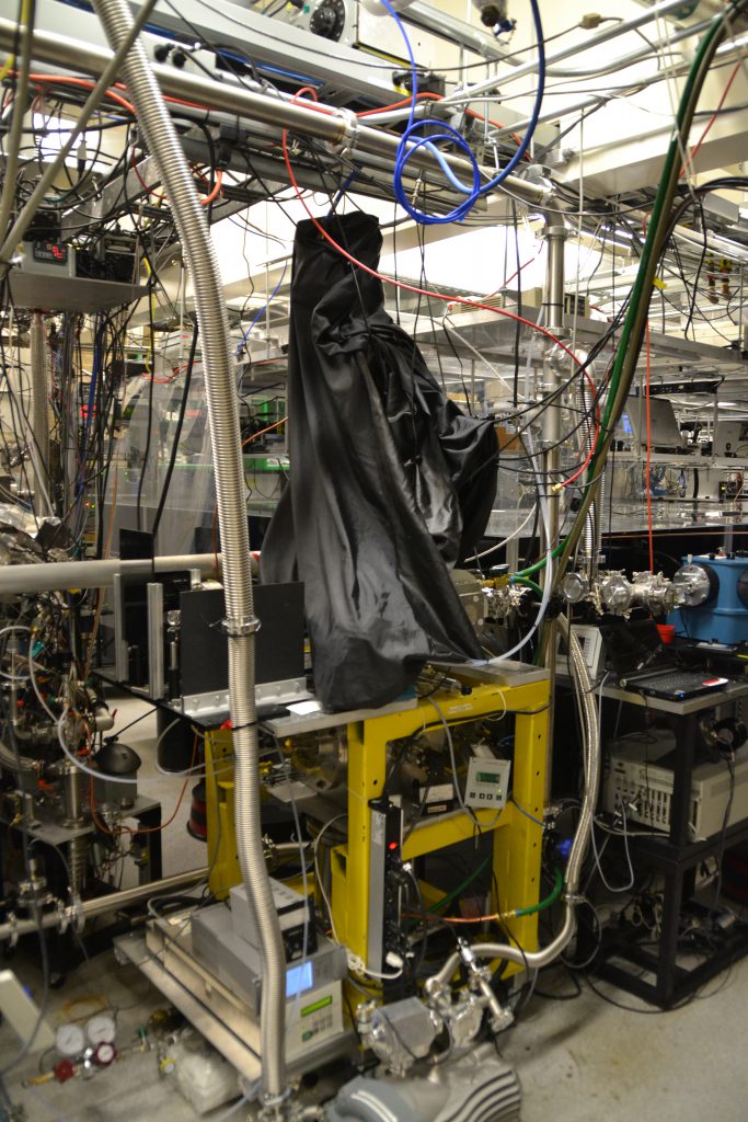
VMI-apparatus 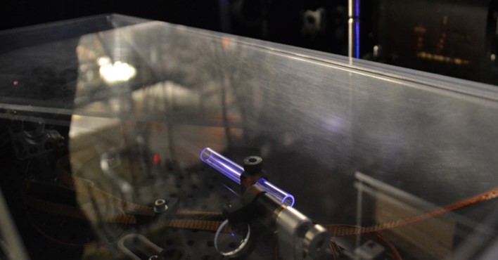
VUV generation